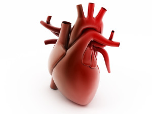
The right ventricle is the heart chamber that pumps blood without oxygen through the lungs via the pulmonary arteries. In patients with PAH, the right ventricle is perhaps the most important barometer or gauge of how things are going. As PAH gets worse, the right ventricle has to work even harder to pump blood through the diseased pulmonary arteries. Eventually if treatments are not effective the right ventricle starts to fail and blood flow to the lungs is reduced and blood backs up into the liver. This leads to lower extremity swelling (edema), abdominal distension, a tender liver and decreased ability to eat a meal.
 Since the right ventricle is so important, we are always trying to figure out ways to best evaluate it’s function. Echocardiography (ultrasound of the heart) is the most common tool used. Much information can be gathered from an echocardiogram such as whether the right ventricle is dilated, how well it squeezes, whether there is fluid around the heart and even a rough estimate of the pulmonary artery pressure. However, because of the shape of the right ventricle precise measurements of the contractility (ability to squeeze) are quite difficult. Many pulmonary hypertension centers now use a measurement called TAPSE (tricuspid annular plane systolic excursion) to obtain more information about the right ventricle’s output from an echocardiogram. This measurement is quite easy to make and provides additional information that has prognostic value. The bigger the number, the better the heart’s function. Normal would be greater than 1.6-1.8cm.
In addition to echocardiography, MRI (magnetic resonance imaging) is emerging as another useful option for evaluating right heart function. The advantages of MRI include the ability to make precise measurements of the right heart dimensions and easy calculation of ejection fraction (squeeze) and stroke volume (amount of blood pumped by the heart on each beat). Furthermore, blood flow measurements can also be made. MRI is particularly good at identifying any abnormal anatomy such as would be found in patients with congenital heart disease. The disadvantages of MRI include that the test takes a while to complete and the patient has to remain perfectly still. The test is also much more expensive than an echocardiogram.
