CT scanning is used during the evaluation process for pulmonary hypertension

A CT of the chest is commonly ordered while evaluating a patient for pulmonary hypertension as it is a useful tool for identifying other lung diseases that might be responsible for the patient’s symptoms. CT scans can be performed as an outpatient at hospitals or imaging centers. There is usually little or no preparation required for a CT scan of the chest but a patient should always check with their physician or imaging center before scheduling the test.
What is a CT scan?
CT scans use x-rays to make detailed images of structures inside the body. The pictures are taken with the patient lying on a table with the CT scanner (a large machine with a hole in the middle) hooked to it. The part of the body being imaged will be in the hole of the scanner. The scanner rotates with each rotation taking less than a second and provides a picture of a thin slice of the area being scanned. All of these pictures are then saved together and the radiologist can view them and move through them as if he is looking directly into the body. This test can be performed with or without contrast. The contrast is an iodine dye that is used to check blood flow and look for other problems. If contrast is ordered an IV will have to be placed and the dye will be injected through the patient’s vein. There is also another variation called an HRCT or high resolution CT scan. This test is the same as a standard CT but takes more pictures of smaller slices of the area of the body being scanned. An HRCT scan can be very useful in evaluating certain types of interstitial lung diseases that may be associated with pulmonary hypertension.
What CT scan results suggest pulmonary hypertension?

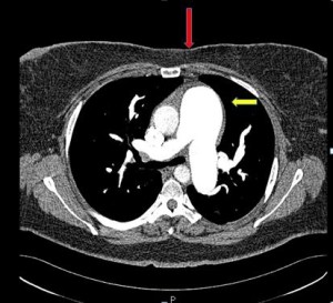
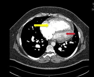
A radiologist will review the images and write a report, which will then be sent to the ordering physician. It is a good idea for the patient to ask for the images to be saved on a disk that they can take with them and show their physician. In most cases no results will be given to the patient at the time of the scan. The CT scan is most useful in diagnosing other lung diseases that may lead to increased pressures in the pulmonary arteries such as blood clots, tumors, or interstitial lung disease (disease in the tissue of the lung not the blood vessels). But findings such as enlarged pulmonary arteries and enlarged chambers of the right heart can be suggestive of pulmonary hypertension. The patient should have a follow up appointment to go over the results with the physician that ordered the test. It is important for the patient to fully understand what the results mean for their treatment plan. It is a good idea for pulmonary hypertension patients to keep a copy of the results for their records.
